Tuesday, 23 December 2014

ECG workshop 2014 | Follow your HEART


 Assalamualaikum wbt and good day everyone!

As a medical student and a future doctor, knowing how to interpret and diagnose the ECG is very important. Learning in the class might be not so enough and need more extra effort to learn it. Therefore, we from IMAM RSC Moscow comes out with the solution to organise the ECG workshop 2014.


Alhamdulillah, all praises go to Allah for His blessing and merciful.

On December 21st 2014, IMAM RSC Moscow was successfully organised the ECG Workshop 2014 that was held at Malaysian Embassy, Moscow Russia.


The event was participated by 60 students form First Moscow State Medical University (FSMU) and Russian State Medical University (RSMU). Most of them are those who are currently in clinical year, where the knowledge about ECG must be great. Insha Allah.

The speaker on that day was Omar Shukri, the 5th year student from First Moscow State Medical University. He presented two wonderful lectures entitled "ECG for beginners" and "How to differentiate?". Next after the break and lunch, the workshop continued with grouping session and quiz. In this grouping session, the participants were divided into 5 small groups and had a discussion about the ECG cases conducted by a few facilitators.


Alhamdulillah, the ECG workshop 2014 finished at 5.00pm with the closing ceremony by the MARA officer in Russia, Encik Saifuddin. Thank you very much to Encik Saifuddin and Puan Fauzana (JPA Officer in Russia) for your time and support.


Last but not least, we would like to thank to Omar Shukri for the great and informative lectures, may Allah count it as your 'amal'. Moreover, a big thank you to the Embassy of Malaysia for giving us the opportunity to organise this such event and not to forget, thank you to all the  participants for coming and support us. We really hope to see you in our next event, Insha Allah.


To the committee members, congratulation! May Allah grant you His rewards and blessings, ameen.


Thursday, 27 November 2014

ECG workshop 2014 | Coming soon

Assalamualaikum wbt and good day guys!

Insha Allah, we are proudly to announce that our ECG Workshop 2014 will be coming soon!

Stay tuned for further information on our facebook page and blog.


see you there!


Monday, 17 November 2014

Pathological ECG findings | ISCHEMIC


Coronary artery disease (CAD)
✔ ️known as atherosclerotic heart disease, atherosclerotic cardiovascular disease, coronary heart disease, ischemic heart disease (IHD), heart attacks.

Myocardial infarction
✔ ️commonly known as a heart attack.
✔ ️An MI occurs when blood stops flowing properly to a part of the heart, and the heart muscle is injured because it is not receiving enough oxygen.
✔ ️Usually this is because one of the coronary arteries that supplies blood to the heart develops a blockage due to an unstable buildup of white blood cells, cholesterol and fat.
✔ ️The event is called "acute" if it is sudden and serious.

ECG Features :

✔ ️ In early STEMIs there may just be peaked T waves with ST elevation developing later.
✔ ️Hyper acute (tall) T waves, ST elevation or new LBBB occur within hours of transmural infarction.
✔ ️ST elevation in two or more adjacent ECG leads.
✔ ️This must be greater than 2 mm (0.2 mV) for males and greater than 1.5 mm (0.15mV) in females if in leads V2 and V3 or greater than 1 mm (0.1 mV) if it is in other ECG leads.
✔ ️ A left bundle branch block that is believed to be new used to be considered the same as ST elevation; however, this is no longer the case.
✔ ️T wave inversion and development of pathological Q waves follow over hours to days

Pathological ECG findings | VENTRICULAR HYPERTROPHY


Left ventricular hypertrophy (LVH)


Definition :
✔ ️the thickening of the myocardium (muscle) of the left ventricle of the heart.
✔ ️LVH itself is not a disease, it is usually a marker for disease involving the heart.
✔ ️Disease processes that can cause LVH include any disease that increases the
afterload that the heart has to contract against, and some primary diseases of the
muscle of the heart.

Causes :
✔ ️aortic stenosis,
✔ ️aortic insufficiency
✔ ️ hypertension.
✔ ️Primary disease of the muscle of the heart that cause LVH are known as hypertrophic cardiomyopathies, which can lead into heart failure.
✔ ️Long-standing mitral insufficiency.


ECG features :

The Sokolow-Lyon index:
S in V1 + R in V5 or V6 (whichever is larger) ≥ 35 mm (≥ 7 large squares)
R in aVL ≥ 11 mm

The Cornell voltage criteria
for the ECG diagnosis of LVH involve measurement of the sum of the R wave in lead aVL and the S wave in lead V3.

The Cornell criteria for LVH are:

S in V3 + R in aVL > 28 mm (men)
S in V3 + R in aVL > 20 mm (women)


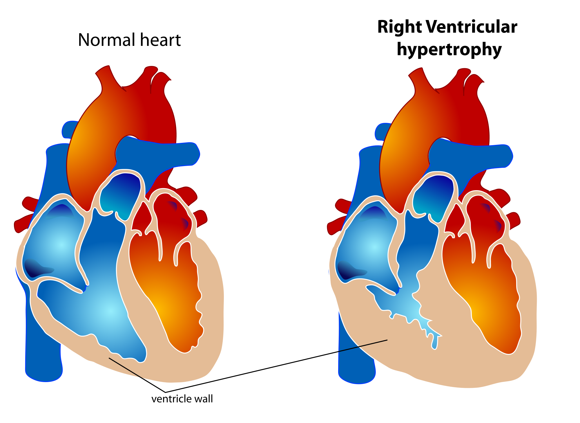
Right ventricular hypertrophy (RVH)


Definition :
✔ ️Right ventricular hypertrophy (RVH) is a form of ventricular hypertrophy affecting the right ventricle.
✔ ️Blood travels through the right ventricle to the lungs via the pulmonary arteries. If conditions occur which decrease pulmonary circulation, meaning blood does not flow well from the heart to the lungs, extra stress can be placed on the right ventricle. This can lead to right ventricular hypertrophy.

Causes :
✔ ️Pulmonary hypertension
✔ ️Tetralogy of Fallot
✔ ️Pulmonary valve stenosis
✔ ️Pulmonic regurgitation
✔ ️Ventricular septal defect (VSD)
✔ ️High altitude
✔ ️Cardiac fibrosis
✔ ️Chronic obstructive pulmonary disease (COPD
✔ ️Athletic heart syndrome


ECG features :

✔ ️Right axis deviation (>90 degrees)
✔ ️Tall R-waves in RV leads; deep S-waves in LV leads
✔ ️Slight increase in QRS duration
✔ ️ST-T changes directed opposite to QRS direction (i.e., wide QRS/T angle)
✔ ️May see incomplete RBBB pattern or qR pattern in V1
✔ ️Evidence of right atrial enlargement (RAE)

Specific ECG features (assumes normal calibration of 1 mV = 10 mm):

✔ ️Any one or more of the following (if QRS duration <0.12 sec):
✔ ️Right axis deviation (>90 degrees) in presence of disease capable of causing RVH
✔ ️R in aVR > 5 mm, or
✔ ️R in aVR > Q in aVR

Any one of the following in lead V1:
R/S ratio > 1 and negative T wave qR pattern
R > 6 mm, or S < 2mm, or rSR' with R' >10 mm

Other chest lead criteria:
R in V1 + S in V5 (or V6) 10 mm
R/S ratio in V5 or V6 < 1
R in V5 or V6 < 5 mm
S in V5 or V6 > 7 mm