What is ECG?
-ECG is used to measure activity of heart,detected by
electrodes attached to the surface of the skin and recorded by a device
external to the body. The recording produced by this noninvasive procedure is termed
an electrocardiogram ( ECG or EKG).
Functions of ECG
- It can give information regarding the rhythm of the heart
- It can also give information regarding the balance of salts (electrolytes) in the blood
- Reveal problems with sodium channels within the heart muscle cells
- Can identify if the heart muscle has been damaged in specific areas, though not all areas of the heart are covered.
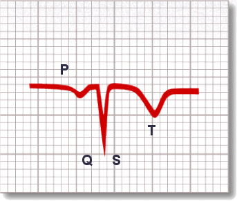
Waveforms of ECG
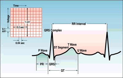
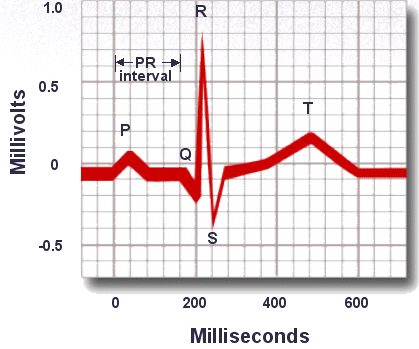
- P wave: depolarization of the right and left atria
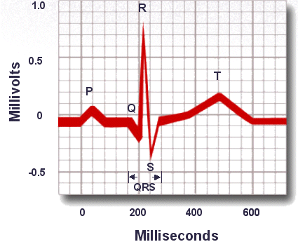
- QRS complex: right and left ventricular depolarization
- ST-T wave: ventricular repolarization
- U wave: probably represents "afterdepolarizations" in the ventricles
- PR interval: time interval from onset of atrial depolarization (P wave) to onset of ventricular depolarization (QRS complex)
- QRS duration: duration of ventricular muscle depolarization due to contraction of this large muscle mass
- QT interval: duration of ventricular depolarization and repolarization
- RR interval: duration of ventricular cardiac cycle (an indicator of ventricular rate)
Here are 10 rules for
normal ECG
Rule 1
PR interval should be 120 to 200 milliseconds or 3 to 5
little squares
Rule 2
The width of the QRS complex should not exceed 110 ms, less
than 3 little squares
Rule 3
The QRS complex should be dominantly upright in leads I and
II
Rule 4
QRS and T waves tend to have the same general direction in
the limb leads
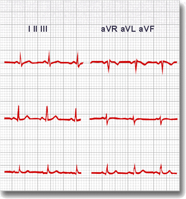
Rule 5
All waves are negative in lead aVR
Rule 6
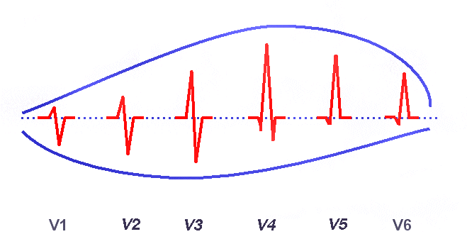
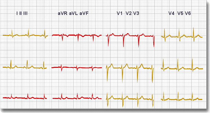
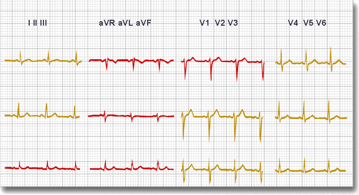
Ø
The R wave in the precordial leads must grow
from V1 to at least V4 (See Below)
Ø
The S wave in the precordial leads must grow
from V1 to at least V3 and disappear in V6. (See Below)
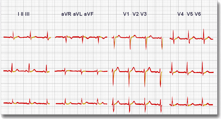
Rule 7
Ø
The ST segment should start isoelectric except
in V1 and V2 where it may be elevated
Rule 8
The P waves should be upright in I, II, and V2 to V6
Rule 9
There should be no Q wave or only a small q less than 0.04
seconds in width in I, II, V2 to V6
Presence of Q wave indicates:
·
Physiologic and positional effects
·
Myocardial injury or replacement MI
·
Ventricular enlargement
·
Altered ventricular conduction
Rule 10
The T wave must be upright in I, II, V2 to V6

Some pathological
changes may be seen on ECG
Ø
Shortened QT interval
Hypercalcemia,
effect of some drugs
Ø
Prolonged QT interval
Hypocalcemia,
some drugs, certain genetic abnormalities
Ø
Flattened or inverted T waves
Coronary
ischemia, hypokalemia, left ventricular hypertrophy, digoxin effect, some drugs
Ø
Hyperacute T waves
Possibly
the first manifestation of acute myocardial infarction, where T waves become
more prominent, symmetrical, and pointed
Ø
Peaked T wave, QRS wide, prolonged PR, QT short
Hyperkalemia,
treat with calcium chloride, glucose and insulin or dialysis
Ø
Prominent U waves
Hypokalemia
Of all the curiosities and common questions encountered on
ecg, we hope that our liltle support page will help you to get there!!
Maybe not to an outstanding result, but once you get a grasp
of the basics, inshaAllah you may find it way easier sooner.
"The knowledge of anything, since all things have
causes, is not acquired or complete unless it is known by its causes."-Ibn
Sina
No comments:
Post a Comment