AV BLOCKS

Definition:
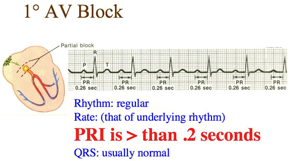
✔ ️A first degree Heart block occurs when electrical impulses moving through the
Atrioventricular (AV) node are delayed (but not blocked). First degree indicates
slowed conduction without missed beats.
ECG Features :
✔ ️Look for rhythm that is regular, with heart rate that is the underlying rate.
✔ ️Notice that the P wave is normal.
✔ ️The PR interval is prolonged (>0.20 sec).
✔ ️The QRS is normal (0.06-0.10 sec).
✔ ️PR Interval is Prolonged (>0.20 sec)
✔ ️QRS is Normal (0.06-0.10 sec)
Notes :
✔ ️A first degree AV block occurs when electrical impulses moving through the
Atrioventricular (AV) node are delayed (but not blocked).
✔ ️First degree indicates slowed conduction without missed beats.

Second degree
1) Second degree Heart Block Type I Wenckebach
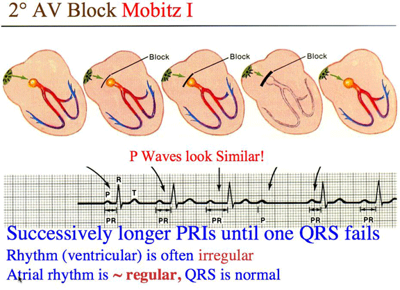
Definition :
✔ ️ it is a condition where the atrioventricular node conducts each successive impulse
earlier and earlier. The PQ interval prolongs from beat to beat up until a drop-out of
one QRS complex. The following impulse will then be conducted normally and the
cycle to begin again. Thus the presence of second-degree AV block is diagnosed
when one or more (but not all) of the atrial impulses fail to conduct to the ventricles.
ECG Features :
✔ ️Look for rhythm that is irregular but with progressively longer PR interval lengthening,
with heart rate that is the underlying rate.
✔ ️Notice that the P wave is normal.
✔ ️The PR interval is progressively longer until a QRS complex is missed, then cycle
repeats.
✔ ️The QRS is normal (0.06-0.10 sec).

2) Second degree Heart Block Type II Mobitz
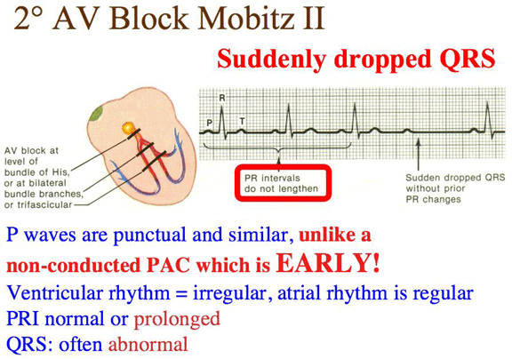
Definition:
✔ ️it is characterized by intermittently non-conducted P waves which are not preceded by
PR prolongation and are not followed by PR shortening.
✔ ️Mobitz II is usually caused by conduction failure/delay of the His-Purkinje system.
✔ ️In three out of four cases, this conduction block is located distal to the Bundle of His,
creating wide QRS complexes.
✔ ️In the other 25% of cases, the block is located within the Bundle of His, producing
narrower QRS complexes.
ECG Features :
✔ ️Look for rhythm that is regular (atrial) and irregular (ventricular), with heart rate that is
characterised by atrial rate usually faster than ventricular rate (usually slow).
✔ ️Notice that the P wave is normal form, but more p waves than QRS complexes.
✔ ️The PR interval is normal or prolonged.
✔ ️The QRS is normal or wide.

Third degree
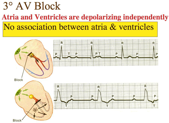
Definition :
✔ ️None of the SA node impulses reach the ventricles.
✔ ️The ventricles will typically compensate by their own pacemaking, known as an escape
rhythm.
✔ ️Thus the atria and ventricles will beat independently and this can be observed on the
ECG.
✔ ️The P waves (atrial beating) and QRS complex (ventricles) are unrelated in time.
✔ ️The PR interval will be variable.
ECG Features :
✔ ️Look for rhythm that is regular, but atrial and ventricular rhythms are independent, with
heart rate that is characterised by atrial rate usually normal and faster than ventricular
rate.
✔ ️Notice that the P wave is normal shape and size, may appear within QRS complexes.
✔ ️The PR interval is absent: the atria and ventricles beat independently.
✔ ️The QRS is normal, but wide if junctional escape focus.

No comments:
Post a Comment