Left ventricular hypertrophy (LVH)

Definition :
✔ ️the thickening of the myocardium (muscle) of the left ventricle of the heart.
✔ ️LVH itself is not a disease, it is usually a marker for disease involving the heart.
✔ ️Disease processes that can cause LVH include any disease that increases the
afterload that the heart has to contract against, and some primary diseases of the
muscle of the heart.
Causes :
✔ ️aortic stenosis,
✔ ️aortic insufficiency
✔ ️ hypertension.
✔ ️Primary disease of the muscle of the heart that cause LVH are known as hypertrophic cardiomyopathies, which can lead into heart failure.
✔ ️Long-standing mitral insufficiency.
ECG features :

The Sokolow-Lyon index:
S in V1 + R in V5 or V6 (whichever is larger) ≥ 35 mm (≥ 7 large squares)
R in aVL ≥ 11 mm
The Cornell voltage criteria
for the ECG diagnosis of LVH involve measurement of the sum of the R wave in lead aVL and the S wave in lead V3.
The Cornell criteria for LVH are:
S in V3 + R in aVL > 28 mm (men)
S in V3 + R in aVL > 20 mm (women)

Right ventricular hypertrophy (RVH)
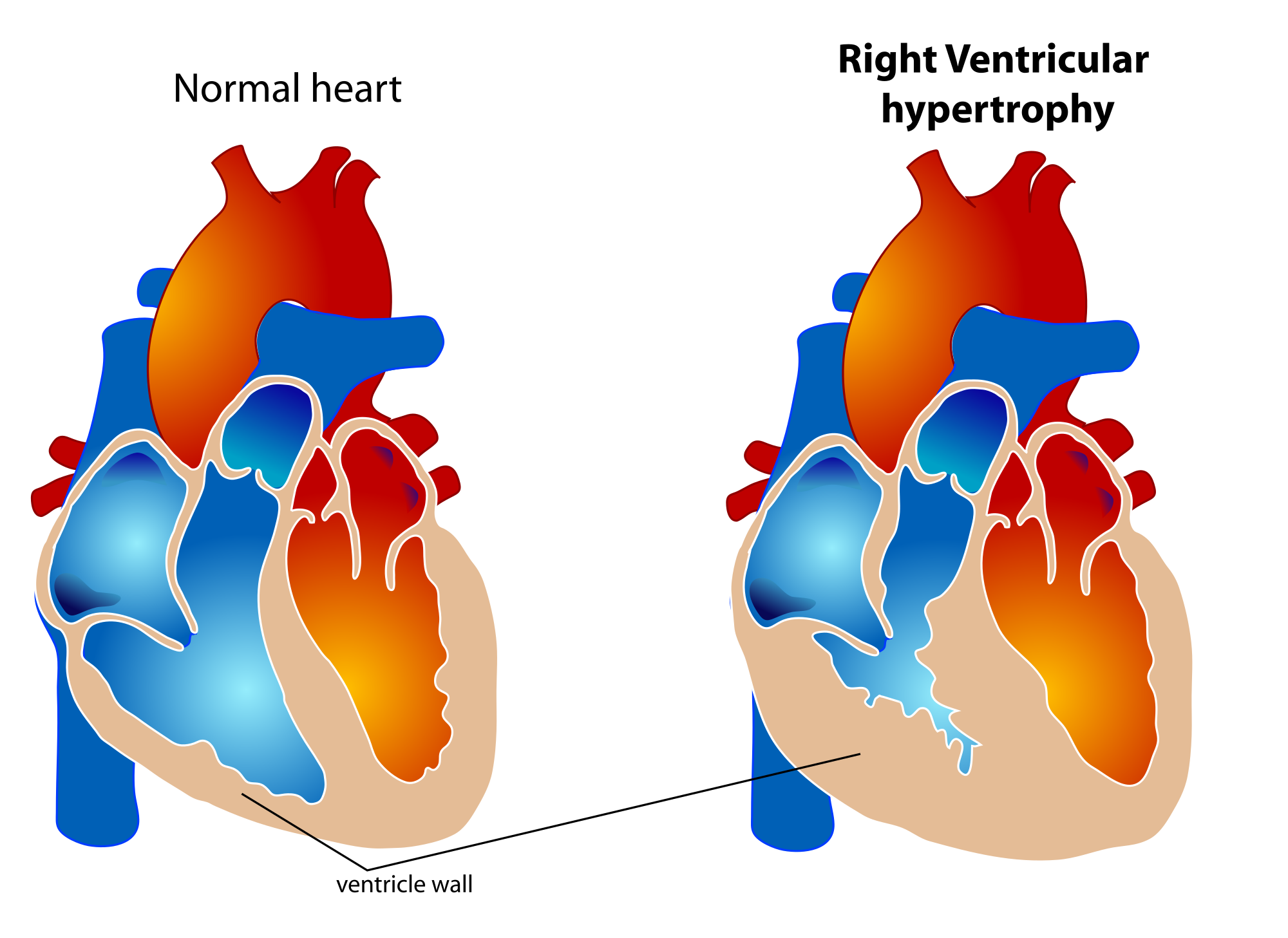
Definition :
✔ ️Right ventricular hypertrophy (RVH) is a form of ventricular hypertrophy affecting the right ventricle.
✔ ️Blood travels through the right ventricle to the lungs via the pulmonary arteries. If conditions occur which decrease pulmonary circulation, meaning blood does not flow well from the heart to the lungs, extra stress can be placed on the right ventricle. This can lead to right ventricular hypertrophy.
Causes :
✔ ️Pulmonary hypertension
✔ ️Tetralogy of Fallot
✔ ️Pulmonary valve stenosis
✔ ️Pulmonic regurgitation
✔ ️Ventricular septal defect (VSD)
✔ ️High altitude
✔ ️Cardiac fibrosis
✔ ️Chronic obstructive pulmonary disease (COPD
✔ ️Athletic heart syndrome
ECG features :

✔ ️Right axis deviation (>90 degrees)
✔ ️Tall R-waves in RV leads; deep S-waves in LV leads
✔ ️Slight increase in QRS duration
✔ ️ST-T changes directed opposite to QRS direction (i.e., wide QRS/T angle)
✔ ️May see incomplete RBBB pattern or qR pattern in V1
✔ ️Evidence of right atrial enlargement (RAE)
Specific ECG features (assumes normal calibration of 1 mV = 10 mm):
✔ ️Any one or more of the following (if QRS duration <0.12 sec):
✔ ️Right axis deviation (>90 degrees) in presence of disease capable of causing RVH
✔ ️R in aVR > 5 mm, or
✔ ️R in aVR > Q in aVR
Any one of the following in lead V1:
R/S ratio > 1 and negative T wave qR pattern
R > 6 mm, or S < 2mm, or rSR' with R' >10 mm
Other chest lead criteria:
R in V1 + S in V5 (or V6) 10 mm
R/S ratio in V5 or V6 < 1
R in V5 or V6 < 5 mm
S in V5 or V6 > 7 mm

No comments:
Post a Comment